Difference between revisions of "KOH/ microscopio"
Geekshabeka (talk | contribs) (→Test Method Instructions) |
|||
| (9 intermediate revisions by 2 users not shown) | |||
| Line 1: | Line 1: | ||
| + | |||
| + | Microscopic observation of unfixed “wet mounts” of | ||
| + | clinical specimens, either stained or unstained, can be useful for the rapid detection of the presence of bacterial, fungal, and parasitic organisms. Presumptive identification can be made, based on morphology and motility. The presence or absence of white blood cells and “clue cells” may also be demonstrated, and a number of well-recognized pathologic conditions may be | ||
| + | identified. | ||
| + | |||
| + | |||
| + | == Test Method Instructions == | ||
| + | |||
| + | |||
| + | '''KOH slide.''' | ||
| + | |||
| + | A sample of the vaginal discharge is placed on a slide and mixed with a solution of potassium hydroxide (KOH)10%.The addition of 10% KOH to the clinical specimen will dissolve tissue cells and keratinized material to allow better visualization of fungal elements.You can replicate the same experiment to detect fungal elements in the skin, for example. | ||
| + | |||
| + | |||
| + | * [[1- Place 1 drop of 10% KOH on the slide.]] | ||
| + | |||
| + | * [[2- Add specimen of vaginal discharge.]] | ||
| + | |||
| + | * [[3- Check the KOH slide immediately for a fishy, amine odor; a strong fishy odor from the mix means vaginosis is present.]] | ||
| + | |||
| + | * [[4- Cover each specimen with a cover slip to exclude air bubbles.]] | ||
| + | |||
| + | * [[5- Yeast/fungi can best be demonstrated on a KOH mount, under DIY/DIWO microscope.]] | ||
| + | |||
| + | * [[6. Examine the KOH preparation under low power (10X) Microscope for yeast pseudohyphae and under high power (40x) for smaller blastospores.]] | ||
| + | |||
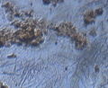
| + | == pre-p-KOH == | ||
| + | |||
<gallery> | <gallery> | ||
| − | |||
File:2015-09-25-225355.jpg | File:2015-09-25-225355.jpg | ||
| Line 16: | Line 43: | ||
</gallery> | </gallery> | ||
| − | |||
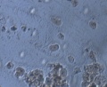
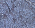
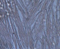
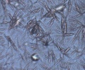
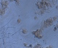
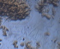
| − | S.PapSmear sin KOH | + | == S.PapSmear sin KOH == |
| + | |||
| + | <gallery> | ||
File:2015-10-03-111501.jpg | File:2015-10-03-111501.jpg | ||
| Line 27: | Line 55: | ||
File:2015-10-03-111722.jpg | File:2015-10-03-111722.jpg | ||
File:2015-10-03-111824.jpg | File:2015-10-03-111824.jpg | ||
| + | File:2015-10-03-115357.jpg | ||
| + | File:2015-10-03-115436.jpg | ||
| + | File:2015-10-03-115548.jpg | ||
</gallery> | </gallery> | ||
| Line 32: | Line 63: | ||
| − | + | ||
| + | |||
| + | [[Category:GynePunk]] | ||
Latest revision as of 23:59, 21 November 2015
Microscopic observation of unfixed “wet mounts” of clinical specimens, either stained or unstained, can be useful for the rapid detection of the presence of bacterial, fungal, and parasitic organisms. Presumptive identification can be made, based on morphology and motility. The presence or absence of white blood cells and “clue cells” may also be demonstrated, and a number of well-recognized pathologic conditions may be identified.
Test Method Instructions
KOH slide.
A sample of the vaginal discharge is placed on a slide and mixed with a solution of potassium hydroxide (KOH)10%.The addition of 10% KOH to the clinical specimen will dissolve tissue cells and keratinized material to allow better visualization of fungal elements.You can replicate the same experiment to detect fungal elements in the skin, for example.
pre-p-KOH
- Error creating thumbnail: File missing