


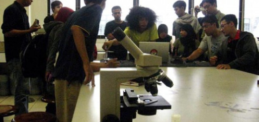

In December 2009, we did an intense discussion session with Gadjah Mada University (UGM) Microbiology lab students. The session is involving both sides on a discussion about developing the DIY microscope from a webcam that has been developed by Hackteria, a platform on open-source biological art, DIY biology and generic lab equipment. Hackteria have given a workshop about DIY webcam microscope in UGM before that was given by Marc Dusseiller (dusjagr). We have built a prototype of the webcam microscope, we asked the students to make tests on this prototype to know its’ limits to used as a tools in analyzing microorganism.
DIY webcam microscope laboratory works
The test started with a pathogen in form of a Systa (a bag of eggs) that normally attacked the roots of potatoes. A normal Systa have a size of 0.5 – 1 mm and could contain 200 – 300 eggs inside of it. This test results to the DIY webcam microscope was able to capture the shape and the movement of the Systa but were unable to capture the structure of the eggs inside of it. The abilities to capture the shape and movement is considered quite helpful for microbiologists as they can analyze the tentacle of that is used by Systa to attached themselves and injects the eggs to the roots of the potatoes.


In the next phase of the test, the students used the DIY webcam microscope to analyze a sample of Saccharomyces cerevisiae, a species that is classified as fungus. S. cerevisiae itself is known since ancient times and thought as the most useful yeast from its use in baking and brewing. S. cerevisiae size is approximately 100 micro (which means it’s size is 100 x 10-6 m) and the DIY webcam microscope still could capture the shape and movement.


Realizing that the lens magnifying capability of the DIY webcam microscope is still far from standard microscope, the session was continued with the use of Hemocytometer device to the test. Hemocytometer is designed for counting cells by placing the samples in to small boxes in the glass and then counted manually by microbiologist with formulas to know the amount of cells that a sample contains. This test in DIY webcam microscope has a very most potential use for Microbiologist as the DIY webcam microscope has several significant advantages than normal analyzing method.


The advantages of the DIY webcam microscope compared to the normal microscope are:
- DIY webcam microscope has a wider scope in the analysis, providing 24 boxes to be viewed in a single analysis. Normal microscope could only capture 4 boxes.
- Normal microscope method is very hard to analyze and need acute precision as it need the researcher to analyze 4 boxes and write manually then slide the Hemocytometer to another 4 boxes without marking the previous one that has been analyzed. DIY webcam microscope could capture and save it as a picture allowing the researcher to count the Hemocytometer in a very significant shorter amount of time.
After the test we develop a discussion to make a shortlist of the improvement that is needed for the DIY webcam microscope prototype. The conclusion of the improvements that is needed are:
- DIY webcam microscope needs an adjustable light intensity. This light intensity is considered very crucial, as the amount of light intensity is very dependable on the focus of the sample.
- Improvements in adjusting lens range to the object sample.
- Lights source from bellow the object sample is needed to get a clearer image of the object.
- A focusing adjustment for the user needed a development to help the user to adjust the slightest movement of the tools.
Research Output & Conclusion
In the next session, we asked the students to give a presentation about Hemocytometer method to further investigate the adjustment needed for the DIY webcam microscope to be used for the method. We wanted to know more about this method and we believe we needed the knowledge to develop the DIY webcam microscope.


They sent two representatives to give a workshop to us about Hemocytometer and they were very enthusiastic to give a presentation to us about it. It seems the last laboratory works in Microbiology creates a motivation for them to develop the DIY Webcam Microscope. As in definition, they said Hemocytometer is a direct method to count the number of microorganism such as bacteria, yeast, spore etc. This method is very important in Microbiology, as Hemocytometer is needed to calculate the concentration of cells in the fluid. According to the students, direct cells counting such as Hemocytometer is very boring process. There are two difficulties that are they encountered in Hemocytometer, which is:
- The moving cells and limited observing area from a microscope is a very difficult process as cells could move from one box to another, making direct counting need a large amount of precision and concentration. It also make direct counting inaccurate.
- To count one sample in Hemocytometer need approximately 10 minutes. However in a laboratory works, scientists must observe hundreds of samples, making it a very long and difficult process.
They stated some of the benefit of using DIY Webcam Microscope, which is:
- Using DIY Webcam Microscope, we can capture photos of sample that is being analyzed and counting the amount of cells at the same time as it captured the whole Hemocytometer.
- DIY Webcam Microscope provides data position in an exact time. Allowing scientists to cross-check the samples. An advantage that couldn’t be done with the standard method of Hemocytometer.
- Very useful for first step analyzing for microscopic object, as it is very useful for shape and morphology analyzing.
We later then continue our session with a discussion. We have to continue on working with the prototype that we developed. During the discussion, lots of idea came to surface relating to the development of the creation. For an example, the quality of lens from the common commercial webcam won’t achieve the same magnification ability such like a normal microscope. We agree that the next step in this collaboration was to optimize the components of the prototype so it has full function in Hemocytometer. To be able to realize the potential that was gain from the laboratory works is set to our next focus. The laboratory works sessions that we did have a good vibe and create mutual understanding on the goal that we wanted to achieve and most importantly how to get there.
The discussion was ended with a short informal session of exchanging information for aesthetic development such as sounds from and affecting microorganism, nanotechnology, wine product research etc. The unlimited possibilities in further collaborative works between us must be seen in both art and science as our background discipline with the limitation of technology surrounding us.
PS3 eye for DIY webcam Hemocytometer
In 2010, dusjagr was giving a second workshop for the Microbiology students in UGM. In this second workshop, dusjagr introduce a DIY webcam microscope from a hacked PS3-eye, a camera that is used for playstation 3 game console. Unlike the generic webcam that we could found in the market, PS3-eye have better image quality and frame rate, enabling the DIY webcam microscope very responsive to slight movement of microorganism.


After the second workshop, Nur Akbar Arofatullah assemble a new prototype using a broken body construction of an analog microscope. The body construction provides accurate precision in driving the lens and the object of observation. This function is obtained to accommodate the shortcomings of the experimental prototype. After going through the re-assembly, Nur Akbar Arofatullah provide a DIY Webcam Microscope for use in the Laboratory of Microbiology Faculty of Agriculture, Gadjah Mada University, Yogyakarta. Until now, DIY Webcam Microscope is always used in the method Hemocytometer by students majoring in microbiology. Calculation results of using DIY Webcam Hemocytometer Microscope is accurate and fit for use in research in the field of microbiology.


Further Development
On 24 November 2010, the work done by the Microfluidics Lab has been publicized as a scientific article in the international journal “Optical Engineering”. Copies of scientific articles that can be obtained by contacting the team on Microfluidics Lab. Current microscope can be operated easily via the USB channel is being used by the Microfluidics Lab to investigate the flow phenomena at the microscopic scale are considered interesting and useful technology.
Research Collaboration Team
- Marc Dusseiller / dusjagr (CH)
- Urs Gaudenz (CH)
- Hackteria.org
- Microbiology lab of Faculty Agriculture Gadjah Mada University, Yogyakarta Indonesia
- Julian Abraham (ID)
- Nur Akbar Arofatullah (ID)
- Bom (ID)
- Subhan Aminun (ID)
- Agus Tri Budiarto (ID)
- Andreas Siagian (ID)
You may read the the same writing in my blog: http://andreassiagian.wordpress.com/2010/03/11/juxtapose-through-media-diy-microscope-webcam-for-hemocytometer-collaborative-research/ or the report in Indonesia on lifepatch site: http://lifepatch.org/Hackteria_-_DIY_Microscope_Webcam


[…] may read the same report in hackteria site: http://hackteria.org/?p=1637 and in bahasa Indonesia on lifepatch site: http://lifepatch.org/Hackteria_-_DIY_Microscope_Webcam […]
[…] and biotechnologies, howeverwhile not up to the standards of professional devices, their DIY microscope webcam for hemocytometer collaborative research is pretty interesting and may have some real world […]
[…] around Akbar, Togar and lifepatch, then continued to do various workshops, both together with UGM and also at various schools and […]